
Hand and Wrist Tumour Removal: What Happens Before a Lump Is Excised?
Learn how hand and wrist lumps are evaluated, when imaging or biopsy may be needed and when surgical removal may be considered.
Most lumps in the hand and wrist are not cancer, but “common” is not the same as “safe to diagnose by appearance.” Ganglion cysts, giant cell tumours of the tendon sheath, fatty growths and several other lesions can produce a visible or palpable mass.
Before removal is planned, the priority is to understand what the lump may be, which structures surround it and whether excision is the correct first procedure.
Does the word tumour mean cancer?
No. A tumour is an abnormal growth of tissue and may be benign or malignant. Many hand masses are benign, while malignant tumours of the hand are uncommon.
The term still deserves careful use. Reassurance should follow an appropriate assessment rather than an assumption that every slow-growing lump is harmless.
Features such as rapid growth, unexplained recurrence, fixation to deeper tissue, neurological changes, skin breakdown or unusual pain can increase concern, but no single feature reliably classifies a mass.
What information helps identify a hand mass?
The history includes when the lump appeared, whether it is changing, previous injury, pain, changes in sensation and how it affects tendon or joint movement.
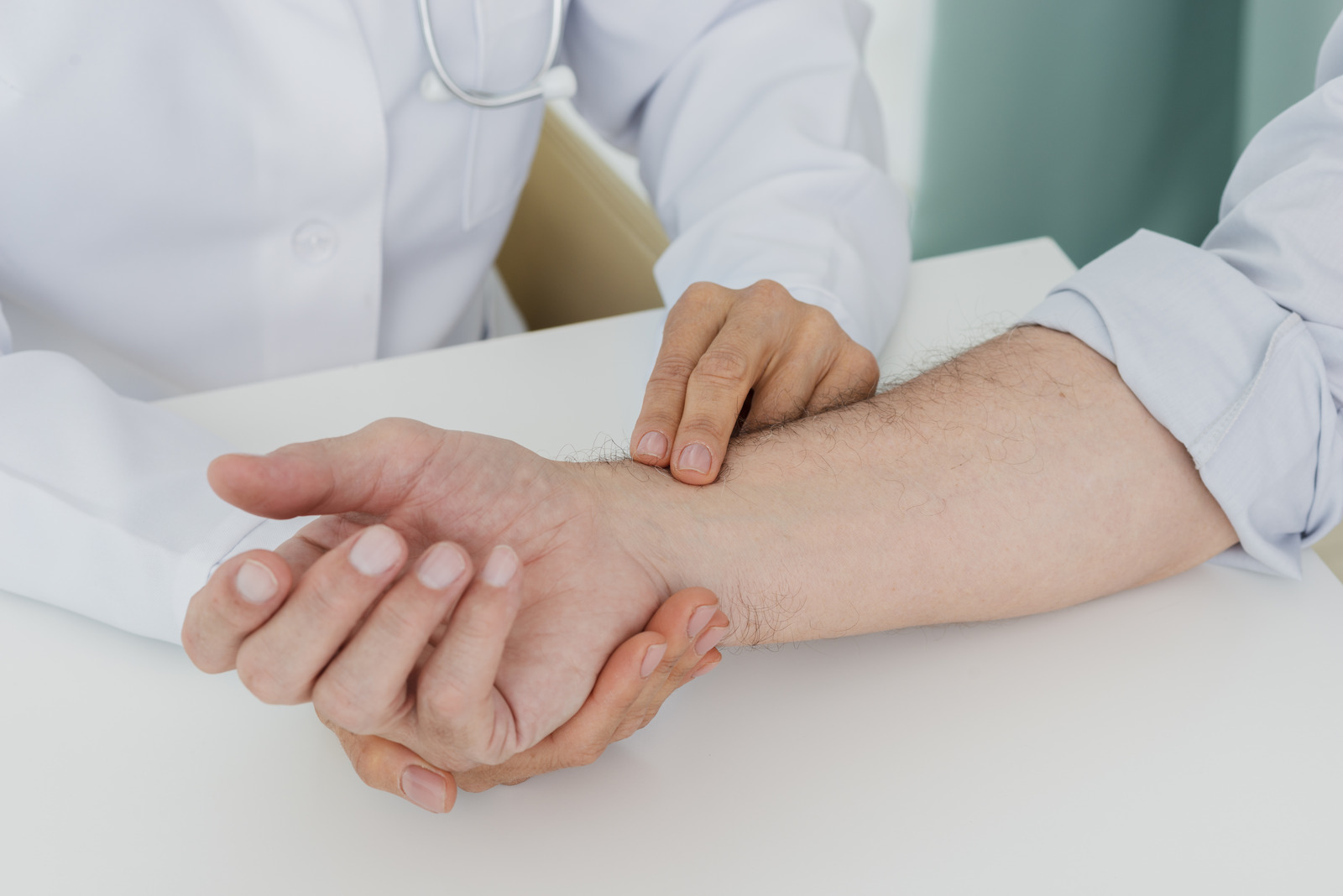
Examination considers location, consistency, mobility, skin changes and the relationship to nerves, blood vessels, tendons and joints. A cystic lump connected to a wrist joint presents differently from a solid mass surrounding a finger tendon.
These entity relationships matter because the hand contains many important structures in a small space. A seemingly minor excision can affect sensation, motion or circulation if the anatomy is not clearly understood.
When is imaging useful?
X-rays can identify bone involvement, calcification, arthritis or changes from an old injury. Ultrasound can help determine whether a lesion is fluid-filled or solid and can show its relationship to a tendon or vessel.
MRI may be considered for a deep, solid, recurrent or poorly defined mass, or when surgical planning requires a more detailed anatomical map. Imaging is selected to answer a question; not every familiar superficial cyst needs every available scan.
If the appearance or imaging raises concern for malignancy, referral and biopsy planning should occur before casual excision. An incorrectly placed biopsy or unplanned removal can complicate definitive treatment.
Is biopsy always performed before removal?
Not always. Some lesions have a sufficiently characteristic presentation that excision and tissue analysis can be combined. Others require a needle or incisional biopsy before the full operation is planned.
The decision depends on size, location, imaging features and the suspected diagnosis. Biopsy is not merely taking a sample from the easiest point; the path and placement need to respect future surgical options.
Any tissue removed is commonly sent for pathological examination so the diagnosis can be confirmed microscopically.
When may surgical removal be considered?
Excision may be appropriate when a benign-appearing mass is painful, growing, compressing a nerve, interfering with tendon movement or joint function, or remains diagnostically uncertain.
The operation should address the lesion while protecting adjacent structures. A mass that wraps around a tendon sheath or lies beside a digital nerve requires a different plan from a superficial cyst.
Removal is not automatically necessary for every stable benign lump. Observation can be reasonable when the diagnosis is sufficiently clear and the mass is not changing or affecting function.
Can a hand tumour return after excision?
Yes. Recurrence depends on the type of lesion, its margins, anatomical constraints and biological behaviour. A benign diagnosis does not always mean zero recurrence risk.
This is another reason pathology matters. Follow-up recommendations for a ganglion cyst, a tendon-sheath tumour and a malignant lesion are not interchangeable.
What risks should be discussed?
Potential risks include infection, scar sensitivity, stiffness, bleeding, injury to a nerve, vessel or tendon, incomplete removal and recurrence. If a mass has already altered nearby structures, removal may not immediately restore normal sensation or motion.
Recovery varies according to depth, location and the amount of reconstruction required. Patients should ask how the procedure may affect work, gripping and postoperative hand therapy.
When does a lump need more urgent assessment?
A rapidly enlarging mass, new weakness or numbness, significant night pain, skin ulceration or a lump returning after previous removal should be assessed without unnecessary delay. Signs of infection—such as spreading redness, warmth, drainage or fever—also require appropriate care.
These findings do not prove cancer, but they make self-diagnosis or indefinite observation less appropriate.
Hand and wrist mass assessment in Montreal
The safest path to removal begins with diagnosis and anatomical planning. The question is not only whether a lump can be excised, but what it is likely to be and what structures must be protected.
If a hand, finger or wrist lump is growing, painful or affecting function, schedule a consultation with Dr. Daniel Durand to discuss examination, imaging and appropriate treatment.